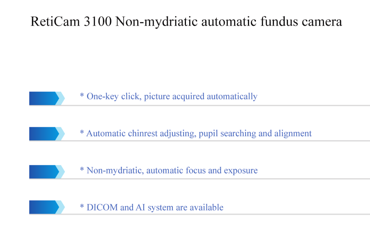
Observed The Color Change Of Optic Nerve Atrophy Digital Fundus
Camera
Digital Fundus camera belongs to the field of medical imaging,
which is used to obtain images of the retina of the human eye, so
that medical personnel can check for fundus diseases or assist
medical personnel to judge the condition of other organs. Because
the blood vessels of the fundus are the only blood vessels that can
be directly observed through the body surface, medical personnel
can check the optic nerve, retina, choroid and refractive medium of
the fundus through fundus camera, and can also help diagnose and
judge other systemic diseases through fundus camera. For example,
through screening retinal photos to detect cerebral infarction,
cerebral haemorrhage, cerebral arteriosclerosis, brain tumors,
diabetes, kidney disease, hypertension, retinopathy of prematurity,
glaucoma, age-related macular degeneration, etc. The earlier these
diseases are detected, the more conducive to clinical treatment.
Therefore, Digital Fundus Camera is widely used in clinical
screening of fundus diseases and has become an indispensable
medical instrument.
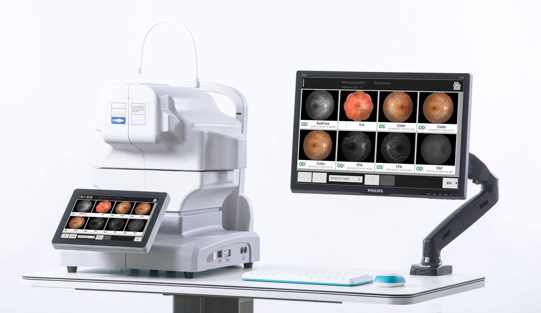
Digital Fundus Camera RetiCam 3100 can be used to capture the
anterior and posterior eye images. It has a field of view of 50
degrees for fundus imaging.
Digital Fundus Camera Reticam 3100 is a highly recognized and
popular automatic fundus imaging system around the world. Through
dual camera imaging, image control and feature recognition, eye XYZ
3D automatic positioning is realized. Through CCD imaging feedback,
the exposure is measured, and the exposure intensity is
automatically and accurately determined, without the need for a
doctor's complex operation. By controlling the focusing step length
according to the image resolution, the image definition can be
automatically adjusted to obtain a clear and accurate fundus image
and help doctors make accurate judgments for patients.
| Acquisition Modes | Digital Fundus Camera/mydriatic Anterior photography
/Red-free(optional)(FFA)/(FAF) |
| Field View | 50° |
| Working Distance | 35mm |
| Minimum Pupil | ≥3.3mm |
| Focus Modes | Manual/Auto |
| Alignment Modes | Double dots auxiliary |
| Exposure | Manual /Auto |
| Photography | SLR Camera |
| Image definition | 24 Megapixel |
| Compensation | ±25D |
| Fixation | External /Internal (any position available) |
| DICOM 3.0 | Support |
| Digital Fundus Camera in the world |
| Brand | TOPCON(NW-400) | DRS | NEWVISON(RETICAM3100) |
| operation | automatic(one key taking picutre)/manual | automatic(one key taking picutre)/manual | automatic(one key taking picutre)/manual |
| Acquisition modes | fundus/anterior/? | fundus/anterior/red-free filter | fundus/anterior/red-free filter/FFA(optional)/FAF(optional) |
| Filed of view | 45 degree | 40-45degree | 45-50degee |
| Mosic degee | | 80 degree | 135degree |
| Minimum pipil size | 3.3mm | 3.5mm | 3.3mm |
| Resolution | above 18megapiexl | sensor size:5 megapixel sensor resoultion: 48 pixel/degree | above18 megapixel (24 megapixel optional ) |
| Fixation | 9 internal + 1 outside fixaiton | 7 internal fixation | countless fixaiton, any position is available during range + 1
outside fixation |
| Technology of focus | | | Dual camrea technology |
| exposure ligh | standard | standard | new technology, more soft, one patient same eye can take picuture
consecutive with almost same performace |

Company Introduction

Focusing on the two major public health problems of adolescent
myopia and elderly ophthalmology, our company researches new
diagnostic and treatment equipment for ophthalmology, develops
low-cost applicable technology products, realizes
industrialization, reduces the cost of social medical and health
system, and serves the strategic needs of national health.The company always adhere to the "high-tech,new vision" for the enterprise development
concept.We have a strong technical force, has been awarded 11 patents, and
obtained 13,485 quality system certification.Bio has established an efficient marketing team and a perfect
after-sales service system to provide medical equipment with the
highest cost performance and meticulous service.


/(FAF) for sale")


